


Onchoceriasis
 There are many types of onchoceriasis. In this project, however, we will focus only on cases of onchoceriasis on humans.
Human onchocerciasis is caused by the filarial parasite Onchocerca volvulus. The infective larvae are normally transmitted by the bite of Simulium flies. Simulium flies can only breed in well oxygenated water because their larvae have an obligatory aquatic stage during which they require high intake of oxygen. Accordingly, onchocerciasis and the blindness it can lead to are associated with fast flowing rivers. As a result, onchocerciasis is often referred to as "river blindness."
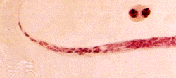
The infective larvae of Onchocerca enter the body through the wound made by the bite of a Simulium fly. The larvae then move through the blood stream to the subcutaneous tissues. In the tissues, they become encapsulated nodules and mature into adults in approximately one year. After mating the female ovivipariously gives birth to microfilariae. The dimensions of microfilariae are about 300 mm in length and 0.8 mm in diameter. Their long and threadlike shape allows them easy navigation through the blood stream. The microfilariae are sheathless with sharply pointed recurved tails.The microfilariae can be found free in the fluid within the nodules and in the dermal layers of the skin. They spread centrifugally from the area where an adult lies. Microfilariae also can be found in the blood and eye during heavy infections. They also infect their fly vectors while the flies are feeding on a host. The microfilariiae then mature into infective larvae in the flies' flight muscles in about ten days.
Of the earliest signs of infection with Onchocerca is the raised nodules that can be seen under the skin around areas over bony prominence. It is suggested that this phenomenon occurs because the larvae are immobilized in these locations while the host is sleeping. The time is long enough for the larvae to be trapped by the body's cellular defense mechanisms. Reactions to dead microfilariae around these nodules can lead to several unpleasant conditions. In the skin there is destruction of the elastic tissues and the formation of redundant folds. There is also often a loss of pigmentation. The histological appearance of advances cases often resemble the skin of very old people. The microfilariae can also enter the eye to cause blindness. Dead microfilariae in the eye often lead to an inflammatory immune response and the eventual formation of secondary cataracts and ocular lesions. As a result, heavy infections often lead to progressive blindness.
The microfilariae can also cause inflammation of regional lymph glands which remove foreign material from the distal skin. This inflammation along with the loss of tissue elasticity can lead to protruding lymph glands enfolded in pockets of skin. This condition is especially prominent in the areas around the scrotum (often called the 'hanging groin' effect) and in severe cases is classified as minor elephantiasis.